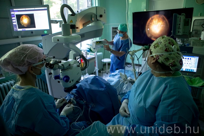
This is the first time the complete system is available in Hungary.
-“Wherever they work, every ophthalmologist wants this microscope.” The lens system, the optical and digital imaging have taken ophthalmic care and education to a new level at the Ophthalmology Clinic. This is the state-of-the art operating microscope in the world – Mariann Fodor; director of the Ophthalmology Clinic of the Clinical Centre (KK) of the University of Debrecen described the new instrument to hirek.unideb.hu.
The microscope equipped with intraoperative OCT is able to accurately show the layers of the retina, enabling the doctor to see much better, much more and finer details during surgery. Patient safety was the most important consideration when purchasing the device.
– The more things and finer details an ophthalmologist sees, the safer the surgery. This is what this microscope facilitates. Better vision shortens surgical time and allows for smaller, more precise manipulations. Use of OCT enables visualisation of the layers of the retina during surgery, greatly contributing to helping a more precise assessment of the processes during the surgery and even immediate monitoring of the effectiveness of the operation, the clinical director explained.
Mariann Fodor added: retinal surgery is an extremely precise and lengthy procedure. During the operation, the ophthalmologist constantly looks down the microscope, struggling with back pain and fatigue by the end of the surgery. The 3D surgical visualization enables the ophthalmologist to perform the procedure with a straight back, operating in special 3D glasses and looking straight ahead at a monitor.
– Digital image processing helps display never before seen details. Anyone who wears 3D glasses while in the operating room can have access to 3D visualization, which creates unprecedented educational opportunities for both colleagues and university students. The most important thing is to give patients the greatest possible chance of complete recovery – the expert of the University of Debrecen explained.
The microscope is used primarily for retinal surgeries, but it also greatly facilitates cataract and corneal surgeries. In addition to full-thickness corneal transplants, it helps separate individual layers of the cornea thinner than a millimetre, making non-full-thickness transplants safer, as doctors can use this tool to determine more accurately which layer they are dealing with during surgery. Use of the tool enables even more complicated cases to be treated more successfully and effectively.
In addition to providing outpatient and inpatient ophthalmic care to more than two million inhabitants of six counties in the Northern Great Plain and Northern Hungary region the Ophthalmology Clinic of the University of Debrecen also has national care responsibilities, including the treatment of malignancies in the eyes. The state-of-the-art tool is also very useful for professionals in this field of diagnostics.
The microscope, valued at more than HUF 150 million, was acquired through the support of the Ministry of Innovation and Technology and with the Ophthalmology Clinic’s own resources. The only place that the best ophthalmic operating microscope system of today is used in Hungary is the Ophthalmology Clinic of the University of Debrecen.
Press Office
unideb.hu


